Over at his blog Oscillatory Thoughts, neuroscientist Bradley Voytek reminds us how we should properly interpret neuroimaging studies to avoid making false claims about "where" in the brain different functions, such as behaviors and mental abilities, are localized: How to be a neuroscientist.
The interpretation of these localization results is confounded, however, by a lack of clarity in what is meant for a "function" to be localized. For example, Young and colleagues (2000) noted that for a given function to be localizable that function "must be capable of being considered both structurally and functionally discrete"; a property that the brain is incapable of assuming due to the intricate, large-scale neuronal interconnectivity.
This is sort of one level higher to what I argued in my first oxytocin post. Just as there is no one gene for any particular property, or indeed no one hormone for any particular property; a particular function is not localized to any particular region of the brain, even though neuroimaging studies sometimes give that impression. Voytek identifies two important principles to go by.
- Behaviors or mental abilities are not discrete/particular definable entities and cannot be "dissected out" and isolated from the whole gamut of behaviors and abilities. I argued the same thing in my second oxytocin post.
- Different regions of the brain interact and interplay, giving feedback to each other and cooperating in very complex ways to produce the brain's functions. Therefore it's unlikely that one particular function, however defined, is localized in any one region of the brain.
Elsewhere Audrey Lustig lists 4 things to keep in mind when you're reading about fMRI:
- Brain pictures of regions "lighting up" (see image below) are often misleading.
- Some fMRI studies are good, some are bad.
- fMRI images don't show what's happening in the brain in real time.
- Neuroscientists can't "read your mind".
Both posts are worth a read!
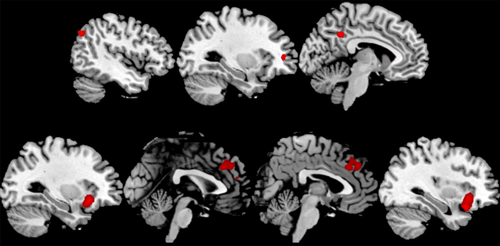
The religious brain compared to the nonreligious brain... see reference here.
Brain imaging studies that study function, especially fMRI, are task dependent and show brain activity during a specific task, subtracting the brain activity from a control state. This means that other brain regions that aren't shown "lighting up" in the brain pictures are active as well during the task, only that they were also active during the control. fMRI reveals differences between one state and the other and if the researchers are not careful about the statistical analysis of their data, they could end up showing the equivalent of brain activity in a dead salmon (original dead salmon fMRI paper).
If you want to see an example of an fMRI study done right, go no longer than to Neuroskeptic and read about Imaging the Brain Better, Faster, Thinner.
Hey, thanks for writing about my post! The neurocritic just blogged about something similar:
ReplyDeletehttp://neurocritic.blogspot.com/2011/02/phrenology-then-and-now.html